


An echocardiogram is a diagnostic test that uses ultrasound to evaluate the heart in real time. It can assess the heart's movement, size, valve structure, heart muscle thickness, overall function, and blood flow.
The test uses high-frequency sound waves to create images of the heart. It can also show major blood vessels connected to the heart, including the pulmonary artery and aorta.
An echocardiogram, also called echocardiography or a heart ultrasound, can be performed using different techniques, including one-dimensional, two-dimensional, and Doppler imaging. A doctor selects the most appropriate type based on what needs to be evaluated.
Main uses
An echocardiogram may be used to:
-
Assess heart function
-
Evaluate the size and thickness of the heart walls, examine the structure of the heart valves, and visualize blood flow
-
Calculate cardiac output, which is the amount of blood the heart pumps per minute
-
Identify changes and diagnose conditions affecting the membrane surrounding the heart, called the pericardium
-
Investigate symptoms such as heart palpitations, shortness of breath, excessive fatigue, or fainting
-
Diagnose heart conditions such as heart murmurs, hypertrophic cardiomyopathy, blood clots in the heart, aneurysms, aortic dissection, tumors, and congenital heart defects
-
Monitor the progression of heart conditions such as heart failure or heart valve disease
An echocardiogram may also be performed to evaluate the heart when coronary artery disease, pulmonary embolism, low blood pressure, respiratory failure of unknown cause, pericardial effusion, or complications from cardiothoracic surgery are suspected.
An echocardiogram is a simple test that can be used to assess heart function in people with or without heart-related symptoms. It may also be used to monitor people with chronic conditions such as high blood pressure or diabetes.
When to get an echocardiogram
There is no set schedule for having an echocardiogram. A cardiologist may recommend the test when there are symptoms, changes in a known heart condition, or another medical reason to evaluate the heart.
When an echocardiogram is used to monitor heart disease, the frequency depends on the type and severity of the condition and whether symptoms or clinical findings have changed.
Routine repeat echocardiograms are generally not recommended for people who are stable, have no symptoms, and have no significant abnormalities that require ongoing monitoring.
Different types
Different types of echocardiograms may be performed depending on what the doctor needs to evaluate. The main types include:
-
Transthoracic echocardiogram: this is the most common type of echocardiogram. The ultrasound transducer is placed on the chest to create images of the heart.
-
Stress echocardiogram: this is performed in a similar way to a transthoracic echocardiogram, but images are taken before and after exercise or the administration of medication in a medical setting.
-
Fetal echocardiogram: this is performed during pregnancy to evaluate the baby's heart and identify possible heart conditions.
-
Doppler echocardiogram: this is especially useful for evaluating blood flow through the heart and assessing heart valve disease.
-
Transesophageal echocardiogram: this test uses an ultrasound probe that is passed into the esophagus to obtain detailed images of the heart.
An echocardiogram may use one-dimensional, two-dimensional, or three-dimensional imaging. Two-dimensional echocardiography provides views of the heart from different angles, while three-dimensional echocardiography creates more detailed, three-dimensional images of cardiac structures.
How to prepare
A standard transthoracic echocardiogram is a simple procedure that generally does not require any special preparation.
A transesophageal echocardiogram requires different preparation. According to the American Heart Association, patients may be instructed not to eat or drink for a certain amount of time before a transesophageal echocardiogram. The healthcare team will provide specific fasting instructions before the test.
Echocardiogram procedure
An echocardiogram is usually performed in a cardiologist's office, hospital, or imaging center and typically takes about 15 to 20 minutes. The person lies on an examination table, usually on their back or left side, and removes or lifts any clothing covering the chest.
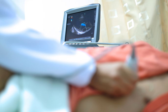
A healthcare professional applies a small amount of gel to the chest and moves an ultrasound transducer over the area. The transducer creates images of the heart from different angles, which are displayed on a monitor.
During the test, the healthcare professional may ask the person to change position or follow specific breathing instructions to obtain clearer images. After a standard echocardiogram, the person can usually return to their normal activities.
Contraindications for use
A standard transthoracic echocardiogram has no specific contraindications and can generally be performed in people of all ages, including infants and children. It may also be performed in athletes and people who are planning to begin exercising.
According to the American Society of Echocardiography, transesophageal echocardiography may be contraindicated in people with conditions such as esophageal narrowing, esophageal tumors, esophageal diverticula, or active upper gastrointestinal bleeding.
A stress echocardiogram may not be appropriate in certain situations, including a recent heart attack, severe symptomatic narrowing of the aortic valve, worsening heart failure, uncontrolled arrhythmias, unstable angina that has not been stabilized, or acute pericarditis.
