


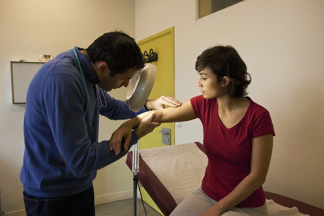
Wood’s lamp, also called Wood light, is a diagnostic tool commonly used in dermatology and aesthetic practice to help detect skin lesions. It also helps assess the type and extent of these lesions based on the fluorescence that appears when the area is exposed to short-wave UV light.
A Wood’s lamp exam is used as a complement to a standard skin exam performed under normal light. It is mainly used to help diagnose skin and nail conditions caused by fungi, and it can also help evaluate porphyria, dark or light skin patches such as vitiligo and melasma, and signs of oily or dry skin.
Based on how the skin looks under UV light, the dermatologist can better identify the lesion and recommend the most appropriate treatment. However, this test does not replace a standard dermatologic evaluation.
Main uses
The main uses of a Wood’s lamp are:
-
Fungal infections, such as tinea capitis or tinea versicolor
-
Bacterial infections, such as erythrasma
-
Acne and post-inflammatory skin pigmentation
-
Pediculosis (lice) and scabies
-
Porphyria
In addition, a Wood’s lamp can be used to assess signs of dryness and excess oil before aesthetic procedures. This helps the professional evaluate the skin’s characteristics and choose the most appropriate treatment.
In pigment disorders, a Wood’s lamp is used not only to assess the borders and characteristics of a lesion, but also to detect subclinical lesions that may not be seen on a routine skin exam and are only visible through fluorescence.
For example, according to the National Institute of Arthritis and Musculoskeletal and Skin Diseases (NIAMS), doctors may use a Wood’s lamp during the evaluation of vitiligo because the ultraviolet (UV) light can make affected areas appear more clearly.
Although a Wood’s lamp is very helpful for diagnosis and follow-up, it does not replace a conventional dermatologic exam.
How it works
A Wood’s lamp is a small device that helps identify different skin lesions based on the fluorescence pattern seen when the area is exposed to short-wave light. Different conditions can appear differently under the lamp, which helps the doctor distinguish one lesion from another.
For the most accurate result, the lesion should be examined with the Wood’s lamp from about 6 inches (15 cm) away, in a dark room without visible light. This allows only the lesion’s fluorescence to be seen during the exam.
Interpreting results
The results of a Wood’s lamp exam vary depending on the color of the fluorescence, which can help the doctor identify and differentiate skin conditions.
On normal skin, the light appears purple without fluorescence, which is considered a negative result.
In infectious skin conditions caused by certain fungi or bacteria, the fluorescence is related to the infectious agent. Based on the MSD Manual, in some bacterial skin conditions such as erythrasma, the affected skin can fluoresce a characteristic coral-red color under a Wood light due to porphyrin produced by the causative bacterium.
In porphyria, fluorescence occurs because of substances present in the urine, which is considered a positive result.
These results should be evaluated by a dermatologist. In some cases, false-positive or false-negative results can occur, especially if the exam room is not dark enough or if the person has used cosmetic products such as perfume, moisturizer, makeup, or hair-bleaching products.