


- A PET scan is an imaging test that helps detect cancer, check for metastases, and evaluate how well treatment is working.
- PET scans use a small amount of radioactive tracer, usually FDG, with a CT scan to show areas with increased metabolic activity.
- Preparation may include fasting, avoiding caffeine and exercise, staying hydrated, and following specific medication instructions.
A PET scan is an imaging test used to investigate some types of cancer. It can help check for metastases, monitor tumor growth, and evaluate response to treatment.
A PET scan is a relatively quick imaging test and is generally considered safe. It uses a small amount of a radioactive tracer (most often a form of glucose called FDG) together with a CT scan to show how metabolically active tumor cells are.
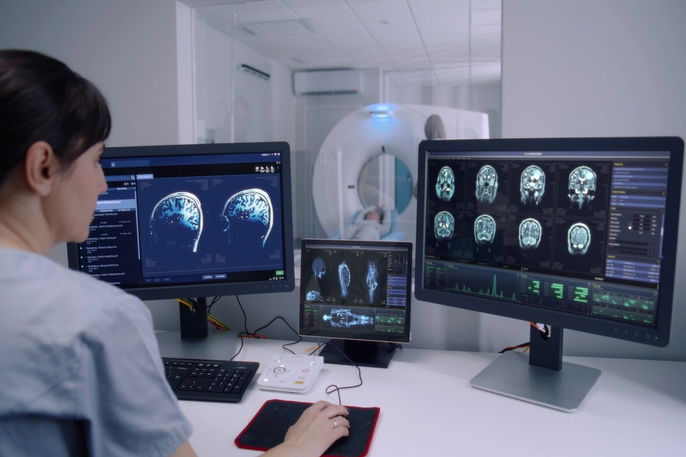
A PET scan is usually performed by a radiologist or a nuclear medicine specialist. The images are then interpreted by a radiologist, and the results are shared with the doctor who ordered the test, such as an oncologist.
What it's for
A PET scan may be recommended in several situations related to cancer care, including:
-
Investigating and diagnosing cancer
-
Assessing whether cancer has spread to other organs (metastasis)
-
Monitoring how the cancer is progressing over time
-
Checking for cancer recurrence or any remaining disease after treatment
-
Determining the stage of cancer
-
Helping estimate prognosis, meaning the likelihood of a favorable or unfavorable outcome
-
Evaluating how well cancer treatment is working
A PET scan is most often used for cancers such as metastatic breast cancer, lung cancer, colorectal cancer, head and neck cancer, esophageal cancer, pancreatic cancer, prostate cancer, thyroid cancer, and brain cancer, as well as melanoma and lymphomas.
According to the National Cancer Institute, imaging tests may be used as part of cancer staging, which helps describe how much cancer is in the body and whether it has spread.
In addition, PET scans can be used to help diagnose neurological conditions, such as epilepsy and dementia, and to evaluate blood flow to the heart, estimate heart attack risk, and determine whether procedures like angioplasty may be needed.
PET scan vs PET-CT
PET scan and PET-CT, or positron emission tomography-computed tomography, refer to the same exam and are simply different names for it.
Both tests use glucose labeled with a radioactive substance and computed tomography to evaluate the metabolic activity of tumor cells.
How to prepare
To prepare for a PET scan, it is recommended to:
-
Avoid physical activity, smoking, and caffeine for 24 hours before the exam.
-
Follow a diet low in carbohydrates and sugar for 24 hours before the exam.
-
Fast completely for at least 6 hours before the exam, or as directed by your doctor.
-
Take your regular medications with a small amount of water up to 1 hour before the exam.
-
Take oral diabetes medications only after the exam. If you use insulin, it may be administered up to 12 hours before the PET scan.
-
Tell the doctor if you have any type of allergy, are pregnant, or think you could be pregnant.
On the day before the PET scan, it is recommended to drink at least 6 to 8 glasses of water throughout the day to stay hydrated.
How it's done
A PET scan is a relatively quick and generally safe imaging test, usually performed in a hospital or outpatient imaging center by a radiology team.
To perform a PET scan, the healthcare professional typically:
-
Injects a small amount of radioactive tracer into a vein, often a form of glucose
-
Waits about 1 hour for the tracer to travel through and be absorbed by the body
-
Asks the person to lie on a table that slides into the scanner
-
Uses the scanner to detect the radiation emitted by the tracer and create detailed images
The PET scan images are then viewed and stored on a computer, allowing the doctor who ordered the test to evaluate the metabolic activity of organs and tissues.
What cancer looks like on a PET scan
When evaluating tumors, for example, cancer cells often use large amounts of glucose because they rely on it as a key energy source to support their growth and division.
As a result, the scan highlights areas that use more glucose, which appear denser and emit more radiation, and these areas may suggest the presence of a tumor.
Aftercare instructions
After a PET scan, you should drink plenty of water and other nonalcoholic fluids and urinate frequently. This helps your body clear the tracer more quickly.
According to SNMMI/EANM guidance, people who are breastfeeding should pause breastfeeding and avoid close contact with infants for 12 hours after FDG administration as a precaution.
Possible side effects
A PET scan is generally considered safe, but it can occasionally cause mild allergic reactions, such as redness at the injection site where the tracer was given.
Main contraindications
A PET scan has few contraindications and can even be performed in people who have diabetes or kidney problems.
People who are pregnant should generally not undergo this exam, as it uses a radioactive substance that may affect the fetus.