


- Angiomas are benign clusters of blood or lymph vessels that can show up as small red to purple bumps on the skin or, less commonly, in the brain or liver.
- Diagnosis may involve a skin exam and, when needed, imaging tests like ultrasound, MRI, or CT; treatment ranges from laser or sclerotherapy to surgery depending on location and symptoms.
- Warning signs require prompt medical attention, especially seizures, sudden or severe headaches, weakness or numbness, trouble speaking, or vision changes that could suggest a brain angioma.
An angioma is an abnormal cluster of blood vessels or lymphatic vessels that can appear on the skin or inside the body. It usually appears as one or more small, soft bumps or spots that range in color from red to purple.
Angiomas are benign (non-cancerous), and the exact cause is not fully understood. Some people are born with an angioma, while others develop one later in life.
Treatment depends on the angioma’s type, size, and location. A dermatologist may recommend laser treatment or sclerotherapy for skin angiomas, while a neurologist or hepatologist (liver specialist) may recommend surgery for angiomas in the brain or liver.
Main symptoms
Angioma symptoms vary depending on where they develop in the body, which include:
1. Skin angioma
The main symptoms of a skin angioma are:
-
A smooth pink or red patch on the face;
-
A small bump, usually red, on the scalp, neck, or trunk;
-
Round red spots with a spider-like appearance;
-
Small red bumps on the skin that can increase in size and number;
-
Bright red, round or oval spots on the skin.
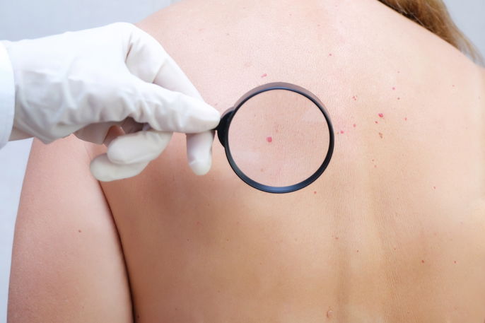
Skin angiomas are the most common type. Although they’re usually harmless, it’s important to have a dermatologist examine them to determine whether any treatment is necessary.
2. Brain angioma
The main symptoms of a brain angioma are:
-
Seizures;
-
Headache;
-
Weakness or numbness;
-
Double vision;
-
Trouble speaking.
A brain angioma can develop in the brain or spinal cord. If you notice any symptoms, see a neurologist as soon as possible to receive the right treatment.
3. Liver angioma
This type of angioma forms on the surface of the liver. It is a small nodule made up of tangled blood vessels, and it usually does not cause symptoms or turn into cancer.
Confirming a diagnosis
A skin angioma is diagnosed by a dermatologist or primary care provider based on your symptoms, medical history, and a physical exam of the skin.
If an angioma is suspected in the brain or liver, a primary care provider, neurologist, or hepatologist may use imaging tests such as ultrasound, MRI, or CT scan to help confirm the diagnosis.
In some cases, a provider may also order a skin biopsy or liver biopsy to confirm the diagnosis and rule out malignant (cancerous) tumors.
Possible causes
The causes of angiomas are not fully known. According to the National Cancer Institute (NCI), benign vascular tumors can form from cells that make blood vessels or lymph vessels and may occur anywhere in the body.
Some factors may contribute to angioma development, such as:
-
Genetic predisposition;
-
Pregnancy;
-
Exposure to certain chemicals, such as bromides or butoxyethanol.
Although some babies are born with an angioma, aging can also increase the risk, especially after age 30.
Types of angioma
The most common types of angioma are:
1. Ruby angioma
Ruby angioma, also called ruby nevus or senile angioma, is a skin angioma that shows up as small red bumps. It typically appears in adulthood and may increase in size and number with age.
2. Cherry angioma
Cherry angioma, also called cherry hemangioma, is another type of senile skin angioma. It mainly affects lighter-skinned adults ages 30 to 50 and usually appears on the arms, trunk, or legs.
3. Strawberry or tuberous angioma
Strawberry (tuberous) angioma is a skin angioma that forms a raised area, usually red. It is often present at birth but can also appear later, growing during the first year of life and then slowly shrinking until it goes away.
4. Spider angioma
This skin angioma has a round, red central spot with small capillaries radiating outward, giving it a spider-like look. It is also called a spider nevus, and its appearance is linked to the hormone estrogen.
5. Flat angioma
Flat angioma, also known as a port-wine stain, is a smooth pink or red patch on the face. It is usually present at birth, but it can also appear months later and often fades after the first year of life.
6. Cavernous angioma
According to the National Institute of Neurological Disorders and Stroke (NINDS), cerebral cavernous malformations (also called cavernous angiomas) are abnormal clusters of closely packed, thin-walled blood vessels that can occur in the brain or spinal cord.
This type is usually congenital (present at birth) but it can occasionally develop later in life and may cause symptoms such as seizures, headaches, or bleeding.
7. Venous angioma
Venous angioma is a type of brain angioma caused by a congenital malformation in certain veins of the brain. These veins are more dilated than normal.
8. Liver angioma
Liver angioma develops on the surface of the liver and usually does not cause symptoms.
The cause of liver hemangioma is unknown, but it is more common in women ages 30 to 50 who have been pregnant or who use hormone replacement therapy (HRT).
Treatment options
Treatment should be recommended by a primary care provider, dermatologist, neurologist, or hepatologist based on the angioma’s size, location, severity, and type.
For skin angiomas, a dermatologist may recommend treatment if the angioma bleeds or causes cosmetic concerns. The main options include:
-
Laser therapy, which reduces blood flow in the vessels and helps remove the angioma;
-
Sclerotherapy, which uses injections to damage and close off blood vessels to help remove the angioma;
-
Electrocoagulation, which uses an electric current delivered through a needle inserted into the angioma to destroy blood vessels and remove it;
-
Cryotherapy, which uses liquid nitrogen spray to help remove the angioma.
These treatments may be used for different types of skin angioma, such as ruby (senile) angioma or spider angioma.
For brain angiomas, treatment should be guided by a neurologist. In some cases, oral corticosteroids such as prednisone tablets may be prescribed to help shrink the angioma.
Surgery may be recommended when a brain angioma is associated with other brain lesions or when a person has symptoms. The goal is to remove the angioma from the brain or spinal cord.
For liver angiomas, treatment is usually not needed because many resolve on their own without health risks. However, if the angioma grows or bleeds, surgery may be necessary.