


- The anatomy scan (often called the 20-week ultrasound) is usually done between 18 and 22 weeks to check fetal growth and major organs, including the brain, heart, kidneys, and spine.
- The scan can help detect certain birth defects and conditions, such as spina bifida, cleft palate, and congenital heart disease, and also evaluates the placenta and amniotic fluid.
- Follow-up imaging may be needed if views are limited or a concern is suspected, and results should be reviewed with an OB/GYN or maternal-fetal medicine specialist.
The anatomy scan is an ultrasound imaging test that lets the healthcare team see the fetus inside the uterus and examine major organs and structures. It can help identify certain conditions or birth defects, such as Down syndrome or congenital heart defects.
This exam is also called a fetal anatomy ultrasound and may be referred to as the 20 week ultrasound. It is most often done in the second trimester between 18 and 22 weeks.
The anatomy scan is a routine part of prenatal care and is often the first opportunity for parents to see detailed images of their developing baby. The results should always be interpreted by an OB/GYN, taking into account the gestational age and overall clinical picture.
What it's used for
The anatomy scan is used to:
-
Confirm gestational age;
-
Assess fetal size by measuring the head, chest, abdomen, and femur;
-
Evaluate fetal growth and development;
-
Monitor the fetal heartbeat;
-
Check the position of the placenta, umbilical cord, and amniotic fluid volume;
-
Detect abnormalities and possible conditions or malformations.
The anatomy scan helps evaluate fetal development and check that growth is progressing as expected.
It can also be used to look closely at organs and structures such as the face, skull, brain, heart, lungs, stomach, abdominal wall, bladder, kidneys, arms, legs, hands, feet, and spine.
If the legs are positioned apart, the clinician may also be able to observe fetal sex, which can later be confirmed with blood testing.
When it's done
The anatomy scan is typically recommended during the second trimester, when the fetus is developed enough for a detailed evaluation. According to the American College of Obstetricians and Gynecologists (ACOG), at least one standard ultrasound is usually performed between 18 and 22 weeks of pregnancy.
This ultrasound can also be performed in the first trimester, between 11 and 13 weeks, but because the fetus is still very small, the images and measurements may be less informative.
In some cases, it may also be done in the third trimester, between 33 and 34 weeks. This is more likely if the scan was not done earlier, if there is concern about a fetal malformation, or if the pregnant person develops an infection that could affect fetal development.
In addition to the anatomy scan, 3D and 4D ultrasounds can show more detail of the baby’s face and can be used when medically indicated to help clarify specific findings. According to ACOG, specialized techniques such as 3D ultrasonography may be used when there is a suspected problem that requires a more detailed evaluation.
Fetal conditions
A second-trimester anatomy scan may help identify several conditions related to fetal development, such as:
-
Spina bifida;
-
Anencephaly or hydrocephalus;
-
Diaphragmatic hernia or gastroschisis;
-
Cleft palate;
-
Kidney abnormalities;
-
Down syndrome, Edwards syndrome, or Patau syndrome;
-
Congenital heart disease.
In this way, the anatomy scan helps evaluate fetal development and can also identify potential structural abnormalities or other health concerns.
How to prepare
In most cases, no special preparation is needed for an anatomy scan.
However, a full bladder can sometimes improve image quality and help lift the uterus. For this reason, the OB provider may recommend drinking water before the exam and avoiding fully emptying the bladder if there is an urge to urinate.
How it's done
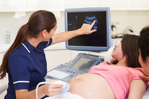
The anatomy scan is typically performed by a trained sonographer in a hospital, imaging center, or obstetric clinic. The images are then reviewed and interpreted by an obstetrician or a maternal-fetal medicine specialist. The exam usually takes about 20 to 60 minutes.
During the exam, the pregnant person lies on an exam table. A thin layer of gel is applied to the abdomen, and the transducer is moved across the skin over the gel.
The device produces images that are displayed on a computer and then interpreted by the clinician.
After the exam, the gel is wiped off with a paper towel, and the person can go home.
Interpreting results
The results of the anatomy scan should be interpreted by an obstetrician or maternal–fetal medicine specialist, taking into account the gestational age and the assessment of key fetal organs and structures, including:
1. Skull and central nervous system
The anatomy scan can evaluate skull and central nervous system development by measuring several parameters, such as:
In addition, the exam may consider the lateral ventricle (LV)/cerebral hemisphere (CH) relationship. This helps assess whether ventricle size is within an expected range and whether ventriculomegaly (enlarged ventricles) is present.
2. Face
Facial measurements may also be evaluated during the anatomy scan, with the main ones including:
In addition, the exam may evaluate the IOD/BPD ratio and the relationship involving the nose/nasal bone.
These facial parameters can help assess changes such as anophthalmia, microphthalmia, and abnormally close-set or wide-set eyes (hypotelorism or hypertelorism), which can be associated with certain syndromes.
3. Abdominal cavity
The abdominal portion of the anatomy scan generally evaluates the following parameters:
The abdominal anatomy scan also allows assessment of organs such as the liver, spleen, and kidneys to check whether they appear to be developing normally or show abnormalities.
4. Thoracic cavity
The anatomy scan may also evaluate parameters of the chest, such as:
This part of the exam may also assess lung volume and chest size, as well as possible lung or heart disease.
5. Bones, spine, and neck
The anatomy scan can evaluate the size and shape of the bones in the legs, arms, hands, and feet, as well as look for signs of bone fractures.
It also evaluates the spine and checks for abnormalities.
The neck and nuchal fold are also assessed, since changes in this area can be associated with certain syndromes, such as Down syndrome.
6. Fetal appendages
Structures that support the fetus are also assessed during the anatomy scan, such as:
In addition to these parameters, the anatomy scan may also be used to estimate the expected due date (EDD).
Also recommended: Gestational Age Calculator: Number of Weeks Pregnant & Due Date tuasaude.com/en/calculate-your-gestational-age