


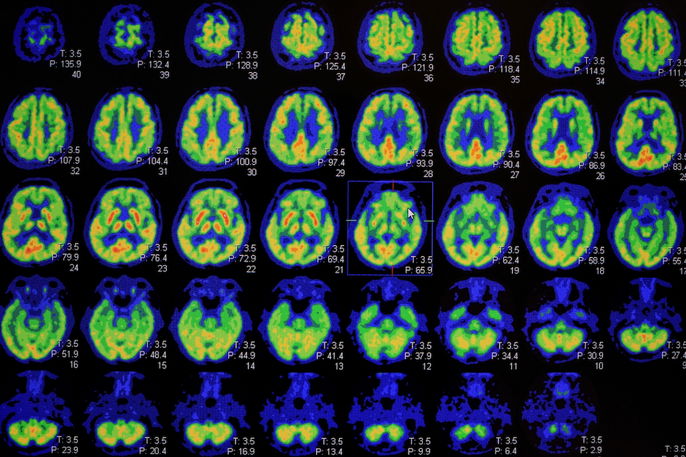
A SPECT scan, also called single-photon emission computed tomography, is an imaging test that helps assess blood flow and activity in the brain. It may be used to help evaluate neurological conditions such as dementia, epilepsy, Parkinson’s disease, stroke, brain tumors, or other disorders.
A SPECT scan may be recommended when other imaging tests, such as an MRI or CT scan, are not enough to confirm a suspected diagnosis. This is because these tests mainly show structural changes in the brain, while SPECT can provide information about how the brain is functioning.
A SPECT scan is done by injecting medications known as radiopharmaceuticals into a vein. These substances can attach to brain tissue and allow images to be created by the scanning device.
Main uses
A SPECT scan provides information about blood perfusion and brain function, and may be useful for:
-
Investigating dementias, such as Alzheimer’s disease or Lewy body dementia
-
Identifying seizure foci in epilepsy
-
Evaluating brain tumors
-
Supporting the diagnosis of Parkinson’s disease or other parkinsonian syndromes, such as Huntington’s disease
-
Assessing neuropsychiatric disorders such as schizophrenia and depression
-
Helping with early diagnosis, monitoring, and progression assessment of cerebrovascular diseases such as stroke and other types of brain hemorrhage
-
Confirming brain death
-
Evaluating traumatic brain injuries, subdural hematomas, abscesses, and vascular malformations
-
Assessing inflammatory conditions, such as herpes encephalitis, lupus, Behçet’s disease, and HIV-associated encephalopathy
A SPECT scan is often ordered when there are doubts about the diagnosis of a neurological condition. According to the Society of Nuclear Medicine and Molecular Imaging (SNMMI), SPECT imaging can evaluate regional blood flow in the brain. This may help identify functional abnormalities that are not visible on structural imaging tests like MRI or CT scans.
Who orders the test
A SPECT scan is usually ordered by a neurologist when there is suspicion of a neurological condition that cannot be confirmed with an MRI or CT scan.
Test procedure
No specific preparation is needed for a SPECT scan. On the day of the test, the patient is usually advised to rest for about 15 to 30 minutes in a quiet room to reduce anxiety and help improve image quality.
Next, the radiopharmaceutical, usually technetium-99m or thallium, is injected into a vein. The patient then waits at least 1 hour for the substance to concentrate properly in the brain before images are taken with the scanner, which usually takes about 40 to 60 minutes. During this time, the patient must lie still without moving, as even small movements can affect the quality of the images.
After the test, patients can resume their normal activities. The radiopharmaceuticals used are generally safe and rarely cause side effects or harm.
Contraindications for use
Brain scintigraphy is contraindicated during pregnancy and lactation. The healthcare provider should be informed if there is any possibility of pregnancy.