


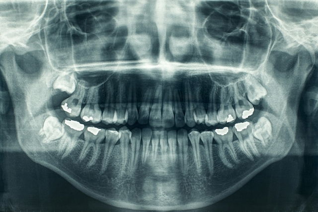
A panoramic X-ray, also called orthopantomography or panoramic radiography, is an imaging test that shows all the bones in the mouth area and their joints, along with all the teeth. It can also show teeth that have not erupted yet, which makes it especially useful for dental treatment planning.
This exam can be performed using either conventional or digital equipment. It allows the dentist to assess tooth alignment, count the number of teeth, and examine the jawbones for issues such as fractures, temporomandibular joint (TMJ) changes, infections, or certain types of tumors.
The radiation exposure from this exam is very low and is not considered a health risk. It is also quick, painless, and can be done in both children and adults.
Indications for use
A panoramic X-ray may be recommended to:
-
Count the number of teeth present in the mouth and within the jaws, such as impacted teeth, and identify missing teeth;
-
Identify the contour of the mandible and assess bone density and the position of the nerve canal for the lower teeth;
-
Observe the amount of remaining bone in periodontal disease;
-
Evaluate the size and shape of the bony structures that form the temporomandibular joints;
-
Identify and assess the maxillary sinuses;
-
Evaluate and compare the shape and size of tooth roots;
-
Provide an overall view of dental health, helping identify cavities and tooth-root abscesses;
-
Check the position of wisdom teeth before extraction;
-
Assess jaw fractures caused by trauma or missing teeth;
-
Evaluate the presence of cysts and tumors in the jaws.
In addition, panoramic radiography may be used to evaluate permanent teeth that are developing in the jaws of children and adolescents before they begin to erupt.
According to the American Dental Association (ADA), dental X-rays are used to help support diagnosis and treatment planning based on each patient’s needs.
Procedure steps
No prior preparation is needed for a panoramic X-ray. The person should remain still during the entire procedure, which is done as follows:
-
A lead apron is worn to protect the body from radiation;
-
All metal objects, such as earrings, necklaces, rings, or piercings, are removed;
-
A plastic lip retractor is placed in the mouth to keep the lips away from the teeth;
-
The face is positioned correctly in the equipment, as directed by the dentist;
-
The machine captures the image, which is then analyzed by the dentist.
After the image is taken, it can usually be viewed within a few minutes, and the dentist can provide a more complete and detailed evaluation of the person’s oral health.
Based on the findings, the dentist may recommend treatments such as a root canal, tooth extraction, restorations, or dental prostheses.
Safety precautions
This exam is considered very safe because it uses only a very small amount of radiation, which is not harmful to your health. However, the American College of Radiology advises anyone who is pregnant (or who might be pregnant) to inform their dentist or oral surgeon, so they can take steps to avoid unnecessary radiation exposure to the fetus.
In addition, people with metal plates in the skull should inform the dentist before undergoing orthopantomography.