


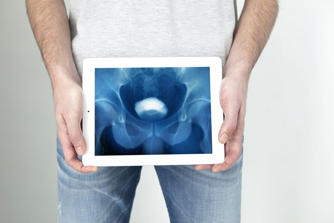
An intravenous pyelogram (IVP), also called excretory urography or intravenous urography (IVU), is an X-ray imaging test that uses IV contrast to assess the structure and function of the urinary system. This includes the kidneys, ureters, and bladder.
This test can help identify changes such as tumors, an enlarged prostate, kidney stones, or hydronephrosis. Hydronephrosis is swelling of the kidney that happens when urine cannot drain into the bladder and builds up in the kidney. This can cause pain, fever, and nausea.
An intravenous pyelogram can take 15 minutes to 4 hours. It is usually performed by a radiologic technologist, and the images are interpreted by a radiologist, a doctor who specializes in imaging tests.
When it's recommended
An intravenous pyelogram is usually recommended to identify changes in the urinary tract, including the bladder, kidneys, and ureters, such as:
- Tumors in the kidneys, bladder, or ureter
- Congenital conditions affecting the urinary system
- Enlarged prostate
- Hydronephrosis
- Scarring caused by urinary tract infections (UTIs)
- Kidney or bladder stones
According to the American College of Radiology, an intravenous pyelogram may be used to evaluate symptoms such as blood in the urine or pain in the side or lower back. It may also be ordered for people with frequent UTIs.
Preparing for the procedure
Preparation for an intravenous pyelogram varies depending on the facility, the reason for the exam, and the person’s age. Preparation may include:
- Not eating or drinking anything after midnight the night before the exam
- Taking a laxative the night before the exam, if recommended by the doctor
People who have difficulty staying still for long periods, such as children or people with claustrophobia, dementia, or schizophrenia, may need sedation during the exam. In these cases, it is important to ask the doctor about any food or drink restrictions before sedation.
How the test is done
During an intravenous pyelogram, the person lies on their back while images of the abdomen are taken. Then, iodinated contrast, a substance that helps make the images clearer, is injected into a vein in the arm or hand, and more images are taken.
During the exam, the technologist may ask the person to empty their bladder so an image can also be taken with the bladder empty.
This exam takes 1 to 4 hours, and it is important to stay still while the images are being taken. The technologist may also ask the person to hold their breath for a few seconds during image capture.
Possible side effects
The contrast injection may cause a slight pinch, irritation, or redness at the injection site. Iodinated contrast can also cause mild symptoms that usually go away after a few minutes, such as flushing, itching, nausea, headache, or a metallic taste in the mouth.
In some people, more serious allergic reactions can occur, leading to shortness of breath, vomiting, or swelling of the mouth or other parts of the body. Based on the American College of Radiology, acute reactions to iodinated contrast may be allergic-like or physiologic and should be managed promptly by the imaging team.
An intravenous pyelogram may not be recommended for people with a known allergy to iodinated contrast.
Potential risks
The risks of an intravenous pyelogram are mainly related to the iodinated contrast injection, which can cause severe allergic reactions in some people. Symptoms may include swelling of the lips and throat, a fast heartbeat, and low blood pressure.
For this reason, any possible allergy symptoms should be reported to the radiology technologist, nurse, or radiologist immediately.