


A HIDA scan is a nuclear medicine imaging test that shows how bile moves through the liver, gallbladder, bile ducts, and small intestine. HIDA stands for hepatobiliary iminodiacetic acid, which is the type of radioactive tracer used during the test.
The scan is mainly used to check the gallbladder and bile ducts. It can help identify inflammation, poor gallbladder emptying, a blocked bile duct, or a bile leak.
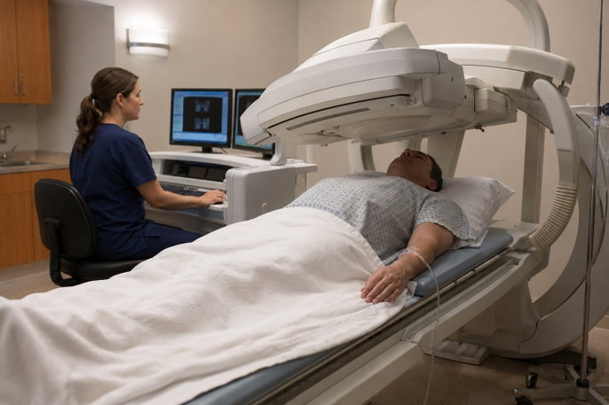
During a HIDA scan, a small amount of tracer is injected into a vein and followed with a special camera. In some cases, medicine or a fatty meal is given to make the gallbladder contract and measure how well it empties.
What a HIDA scan is used for
A HIDA scan may be ordered when symptoms or other imaging tests suggest a problem with the gallbladder or bile ducts. Common reasons for the test include:
-
Acute cholecystitis, which is sudden inflammation of the gallbladder
-
Chronic cholecystitis, which is long-term gallbladder inflammation
-
Biliary dyskinesia, in which the gallbladder does not empty normally
-
A blocked bile duct
-
A bile leak, especially after surgery or an injury
-
Certain problems involving the sphincter of Oddi, a muscle that controls the flow of bile into the small intestine
The test may be especially useful when an ultrasound does not clearly explain symptoms. For suspected acute cholecystitis, failure of the gallbladder to appear during the scan may indicate that the cystic duct is blocked.
How the HIDA scan is performed
The test is performed in a nuclear medicine department. A small intravenous, or IV, line is placed in a vein, usually in the arm or hand.
A small amount of radioactive tracer is injected through the IV. The tracer travels through the bloodstream to the liver, where it is removed from the blood and released into the bile.
The tracer should then move through the bile ducts, enter the gallbladder, and pass into the small intestine. A gamma camera positioned over the abdomen records images as the tracer moves through these organs.
It is important to remain as still as possible while the images are taken. The test commonly lasts between one and several hours, depending on how quickly the tracer moves and whether extra imaging is needed.
Normal HIDA scan results
A normal HIDA scan generally shows that the tracer:
-
Is taken up by the liver
-
Enters the bile ducts
-
Reaches the gallbladder within the expected period
-
Passes into the small intestine
-
Remains within the liver and biliary system without leaking into nearby tissues
When gallbladder emptying is measured, the report may include a gallbladder ejection fraction, also called GBEF. This percentage shows how much bile leaves the gallbladder after stimulation with sincalide or a fatty meal.
What a high ejection fraction means
A high gallbladder ejection fraction means the gallbladder emptied a large percentage of its contents during the test. There is no widely accepted cutoff that defines a high result in all patients and protocols.
An unusually high value may be described as gallbladder hyperkinesia or a hyperkinetic gallbladder. This means the gallbladder contracts very strongly or empties very quickly after stimulation.
Some people with a high ejection fraction have pain that resembles gallbladder pain. However, the clinical meaning of a high result remains uncertain, and research has not established that a high ejection fraction alone confirms gallbladder disease.
A high result should therefore be considered together with symptoms, whether pain occurred during gallbladder stimulation, and whether other causes of abdominal pain have been ruled out.
What a low ejection fraction means
A low gallbladder ejection fraction means the gallbladder released less bile than expected after stimulation. Depending on the protocol, values below approximately 35% to 38% may be considered low.
A low result may suggest gallbladder hypokinesia, meaning reduced gallbladder movement. It is commonly associated with biliary dyskinesia or functional gallbladder disorder.
Possible causes or related conditions include:
-
Poor gallbladder muscle contraction
-
Chronic gallbladder inflammation
-
Functional gallbladder disease
-
Certain medicines, especially opioid pain medicines
-
An unsuitable fasting period
-
Differences in how sincalide or a fatty meal was given
A low ejection fraction does not always prove that the gallbladder is the cause of pain. Results may vary between testing methods, and some people with low values do not improve after gallbladder surgery.
Other abnormal HIDA scan results
The ejection fraction is only one part of the test. The timing and location of the tracer can reveal other problems.
1. Gallbladder not seen
When the gallbladder does not fill with tracer, the cystic duct may be blocked. Persistent nonvisualization of the gallbladder is an important finding in acute cholecystitis.
Delayed gallbladder filling may be associated with chronic gallbladder disease, although the full scan pattern and clinical findings are needed for interpretation.
2. Tracer does not reach the intestine
Delayed or absent movement of tracer into the small intestine may suggest a bile duct obstruction. The pattern may differ depending on whether the blockage is partial or complete.
Poor liver function can also slow tracer movement. For this reason, delayed passage does not always mean that a bile duct is blocked.
3. Tracer appears outside the bile ducts
Tracer found outside the expected biliary system may indicate a bile leak. This can occur after gallbladder surgery, liver surgery, transplantation, injury, or another procedure involving the bile ducts.