


- A breast ultrasound helps evaluate breast lumps, cysts, inflammatory changes, and abnormalities found on a breast exam.
- This test can complement mammography, especially in people with dense breast tissue or higher breast cancer risk.
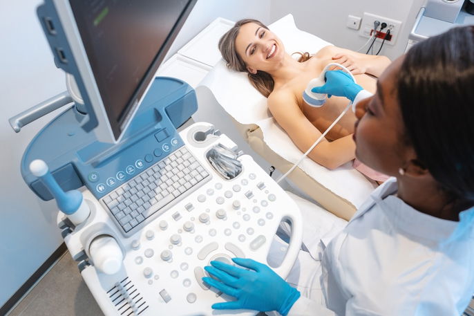
- Breast ultrasound is quick and painless, and the images are reviewed by a radiologist using the BI-RADS classification.
A breast ultrasound is an imaging test used to evaluate the internal structures of the breast. It can help identify breast lumps, breast cysts, breast cancer, or inflammatory changes, for example.
This test is usually ordered when a breast abnormality is found on a mammogram. It can also be used to guide a breast biopsy.
Breast ultrasound is a simple, quick, and painless test. No preparation is needed, although the person will usually be asked to remove their shirt and bra, when applicable.
What it is used for
A breast ultrasound is used to evaluate breast abnormalities found during a breast exam, mammogram, or MRI, such as benign cysts, solid lumps, or breast cancer.
According to the American College of Radiology, supplemental screening may be considered based on a person’s breast density and individual breast cancer risk, especially in people with very dense breasts.
Breast ultrasound may also be used to guide a breast biopsy, helping the doctor locate the breast lump so a needle can be inserted to collect a sample from the lesion.
When it is recommended
Breast ultrasound may be recommended to:
-
Differentiate between benign and malignant lumps
-
Detect seromas and hematomas
-
Check the condition of breast implants
-
Investigate the cause of breast pain or inflammatory changes
-
Assess the characteristics of palpable lumps
-
Monitor lumps
Breast ultrasound may also be recommended for people who are pregnant or at high risk of breast cancer who cannot be exposed to radiation from a mammogram or MRI.
The doctor may also recommend a breast ultrasound to evaluate how well chemotherapy and/or radiation therapy is working to treat breast cancer.
Breast ultrasound vs mammogram
A breast ultrasound is not the same as a mammogram and does not replace this test. It is only used to complement breast evaluation.
Mammography is still the most recommended test for identifying breast cancer.
What to expect
A breast ultrasound is usually performed by a trained sonographer in specialized clinics or hospitals, and the images are interpreted by a radiologist. It is a test that does not cause pain or discomfort.
To perform a breast ultrasound, the sonographer usually follows these steps:
-
Asks the person to remove their shirt and bra, when applicable, and put on a gown that opens in the front
-
Asks the person to lie on the exam table with one arm positioned behind the head
-
Applies gel to the ultrasound transducer and to the breast being examined
-
Places the transducer on the skin and moves it over the breasts
- Checks the images shown on the computer during the exam
- Saves images for the radiologist to review and include in the ultrasound report
During the ultrasound, the sonographer captures images that allow the radiologist to assess the characteristics of a lump or cyst in the breast, such as whether it is solid or filled with fluid, its shape, and whether its borders are well-defined or poorly defined.
Based on the American College of Radiology’s BI-RADS system, breast ultrasound results are organized into standardized categories that help radiologists communicate findings and guide follow-up recommendations. This system divides findings into categories to help determine the risk of breast cancer.
Automated breast ultrasound
Automated breast ultrasound, or ABUS, is a type of breast ultrasound performed with a device that has a larger transducer and automatically scans the breast. It is commonly used as a supplemental screening test for people with dense breast tissue.
Although ABUS scans nearly the entire breast and generates hundreds of images, a traditional handheld breast ultrasound may still be needed if an abnormality is found.
Interpreting results
Breast ultrasound results are reported according to the BI-RADS classification:
Breast ultrasound results should be interpreted by a breast specialist, gynecologist, or oncologist, together with the mammogram and/or breast MRI.