


A brain CT scan is a diagnostic imaging test that uses X-rays to create images of the brain, skull bones, neck, ears, eye sockets, and sinuses. This brain scan is commonly used to investigate symptoms such as headaches, seizures, dizziness, muscle weakness, sudden changes in behavior, hearing or vision loss, and difficulty speaking or swallowing.
The test can help doctors diagnose several health conditions, including stroke, brain aneurysm, brain cancer or tumors, epilepsy, meningitis, hydrocephalus, and Alzheimer’s disease. It may also be used to help plan radiation therapy for the brain.
A brain CT scan can be performed with or without radiologic contrast. The results should be evaluated by a neurologist.
Why it is ordered
A brain CT scan may be used to evaluate conditions that can affect the head and neck, such as:
- Stroke;
- Brain aneurysm;
- Cerebral thrombosis;
- Encephalomalacia;
- Brain hemorrhage or bleeding;
- Cerebral edema;
- Brain cancer or tumors;
- Alzheimer’s disease.
In addition, a brain CT scan can help diagnose Parkinson’s disease, multiple sclerosis, brain tissue atrophy, epilepsy, meningitis, head trauma, injuries to the face, head, or skull, hydrocephalus, skull diseases or malformations, and arteriovenous malformations.
A brain CT scan may also be used to guide needle placement during a brain biopsy or to help plan radiation therapy for brain cancer.
How it is done
A brain CT scan is a quick, painless test that takes an average of 10 to 30 minutes and is performed by a radiologist or neurologist.
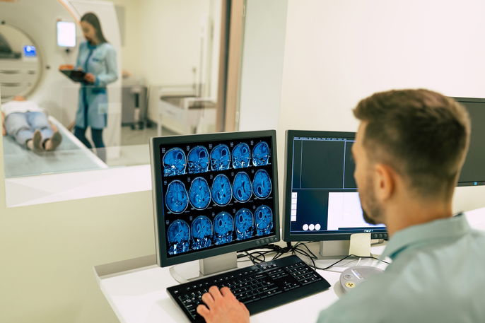
For the test, the person lies on their back on the table of the CT scanner. The scanner is shaped like a ring and emits X-rays that pass through the skull and are processed by a computer to create images of the head, which are then evaluated by the doctor.
During the test, the person needs to remain still so that movement does not affect the results. The images are processed and stored as the scan is performed, and children may need anesthesia.
Brain CT scan with contrast
A brain CT scan with contrast is performed to evaluate the blood vessels in the brain.
Before a CT scan with contrast, the doctor may administer saline solution through a vein in the hand or arm. An iodinated contrast agent is then injected into the vein to provide clearer images and allow better evaluation of the blood vessels.
This test supplements the initial evaluation performed without contrast and provides clearer images.
How to prepare
Some precautions may be needed when preparing for a brain CT scan, such as:
- Tell the doctor about any allergy to iodine or any type of radiologic contrast, latex, medications such as penicillins, or foods such as shellfish or seafood before the test;
- Tell the doctor about any oral diabetes medications being used, such as metformin, before the test, as the doctor may recommend temporarily stopping the medication around the time of a CT scan with contrast;
- Bring a list of all medications, vitamins, and dietary supplements that are taken regularly;
- Tell the doctor about any health conditions, such as hyperthyroidism or kidney failure;
- Fast for about 4 hours before a CT scan with contrast;
- Take regular medications as usual with a small amount of water, as directed by the doctor;
- Do not wear earrings, a watch, or other jewelry on the day of the test.
The doctor should also be informed if the person has kidney problems or uses a pacemaker or another implanted device.
Care after the scan
After a brain CT scan with contrast, plenty of water or other nonalcoholic fluids should be consumed. Frequent urination can help eliminate the remaining contrast agent.
MRI vs. CT Scan: Which Is Better for the Brain?
CT scans are generally better at spatial resolution, showing sharp edges between structures, while MRI is better at contrast resolution, distinguishing between different types of soft tissue.
In practice: CT is usually first in emergencies, and MRI is usually better for detailed, non-urgent evaluation.
Contraindications
A brain CT scan should not be performed if pregnancy is suspected or confirmed.
In addition, iodinated contrast should not be used in people who are allergic to iodine or have kidney failure.
Possible complications
A brain CT scan is generally considered safe and usually does not cause complications during the procedure. However, in rare cases, allergic or anaphylactic reactions to iodinated contrast may occur.
For this reason, a brain CT scan should be performed by a radiologist in a specialized imaging center or hospital that has equipment and staff available to provide immediate emergency care if a complication occurs.