


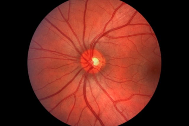
Hypertensive retinopathy refers to changes in the back of the eye caused by high blood pressure. These changes can affect the arteries, veins, and nerve tissue in the retina.
The retina is the light-sensitive layer at the back of the eye. It converts light into nerve signals that the brain uses to create visual images.
Although the condition mainly affects the retina, high blood pressure can also damage the choroid and optic nerve. Hypertensive retinopathy may develop gradually in people with long-term high blood pressure or appear suddenly during a severe spike in blood pressure.
Main types and symptoms
Hypertensive retinopathy may be chronic when it is associated with long-term high blood pressure, or malignant when it is associated with malignant hypertension:
1. Chronic hypertensive retinopathy
Chronic hypertensive retinopathy usually does not cause symptoms and develops in people who have had high blood pressure for a long time. It can cause the small blood vessels in the retina to become narrow, change how they reflect light, and alter the way arteries and veins cross over each other.
In less common cases, retinal hemorrhages, microaneurysms, and signs of blocked blood vessels may also occur.
2. Malignant hypertensive retinopathy
Malignant hypertensive retinopathy is associated with a sudden increase in blood pressure, with systolic blood pressure above 200 mmHg and diastolic blood pressure above 140 mmHg. This condition can affect not only the eyes but also the heart, kidneys, and brain.
Unlike chronic hypertensive retinopathy, which is usually asymptomatic, malignant hypertensive retinopathy commonly causes headache, blurred vision, double vision, and a dark spot in the field of vision.
It may also cause changes in retinal pigmentation, macular edema, detachment of the neurosensory retina in the macular area, and ischemic optic disc swelling with retinal hemorrhages and cotton-wool spots.
Confirming a diagnosis
Hypertensive retinopathy is diagnosed with fundoscopy, also known as a fundoscopic examination. During this exam, an ophthalmologist uses an instrument called an ophthalmoscope to examine the back of the eye and the structures of the retina for changes that may affect vision.
Fluorescein angiography may also be performed. According to the American Academy of Ophthalmology (AAO), this type of retinal imaging may be used when the diagnosis is uncertain, to assess retinal blood vessel damage, or to rule out other eye conditions.
Grades of hypertensive retinopathy
Hypertensive retinopathy caused by high blood pressure is classified into the following grades:
-
Grade 0: no visible changes
-
Grade 1: moderate narrowing of the retinal arterioles
-
Grade 2: marked narrowing of the retinal arterioles with focal irregularities
-
Grade 3: the same changes as Grade 2, along with retinal hemorrhages and/or exudates
-
Grade 4: the same changes as Grade 3, along with optic disc swelling
Treatment options
Chronic hypertensive retinopathy usually does not require specific eye treatment. However, treatment by an ophthalmologist may be needed if complications affect the retina.
Malignant hypertensive retinopathy is a medical emergency. According to the American Heart Association (AHA), hypertensive emergencies require immediate, carefully controlled blood pressure reduction to prevent or limit permanent organ damage.
Once the hypertensive crisis is treated, vision may improve partially or return completely.