


Cranial nerves are 12 pairs of nerves that connect the brain with structures in the head and neck. They control functions such as smell, vision, hearing, facial movement, and certain automatic body processes.
These nerves can carry sensory information to the brain, including visual, auditory, smell, and facial sensations. They may also transmit motor signals that control the eyes, face, tongue, jaw, and internal organs, while some cranial nerves perform both sensory and motor functions.
A cranial nerve assessment can help identify nervous system abnormalities through movement and reflex tests, such as the pupillary and corneal reflexes. It can also help determine the location of damage affecting the central or peripheral nervous system.
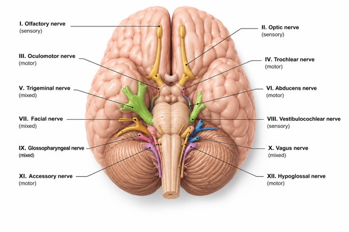
The 12 pairs of cranial nerves and their functions are:
1. Olfactory nerve (I)
The olfactory nerve, also known as cranial nerve I, is a sensory nerve made up of neurons located in the upper part of the nose, in an area called the olfactory mucosa.
Functions: The olfactory nerve allows the brain to detect smells through specialized cells in the upper part of the nose. These cells identify odors and send electrical signals to the olfactory bulb.
The brain then receives and interprets these signals, allowing it to recognize and distinguish different smells.
If this nerve is damaged, signals may not reach the brain properly. This can lead to a loss of smell, known as anosmia, or changes in the perception of odors, known as parosmia or dysosmia.
2. Optic nerve (II)
The optic nerve, also known as cranial nerve II, begins in the retina and carries nerve impulses to an area of the brain called the thalamus.
Functions: The optic nerve carries visual information from the retina to the brain.
In the retina, rods and cones detect light and convert it into electrical signals, which are then transmitted to ganglion cells. The axons of these cells come together to form the optic nerve, which carries the information to the brain.
The brain interprets these signals to recognize shapes, colors, and movement. It also helps regulate the pupils’ response to light.
If the optic nerve is damaged, visual signals may not reach the brain correctly. This can cause vision loss, visual field defects, or difficulty distinguishing colors.
3. Oculomotor nerve (III)
The oculomotor nerve, also known as cranial nerve III, originates in the brainstem and connects mainly to muscles within the eye socket.
Functions: The oculomotor nerve is a motor nerve that controls most eye movements and helps the pupil adjust to light.
This nerve sends signals to most of the muscles responsible for moving the eyes and raising the upper eyelid.
It also allows the brain to control pupil constriction, which can be observed when light enters the eyes.
If this nerve is damaged, eye movement may be impaired. This can cause double vision, a drooping eyelid, or dilated pupils that do not respond to light.
4. Trochlear nerve (IV)
The trochlear nerve, also known as cranial nerve IV, helps control eye movement and works together with cranial nerves III and VI.
Functions: The trochlear nerve is a motor nerve that allows the eye to move downward and inward. It also helps rotate the eye inward during activities such as reading or walking down stairs.
If the trochlear nerve is damaged, it may cause double vision, especially when looking downward and outward.
5. Trigeminal nerve (V)
The trigeminal nerve, also known as cranial nerve V, divides into smaller branches that extend throughout the face, particularly to the skin and certain muscles in the jaw.
Functions: The trigeminal nerve is a mixed sensory and motor nerve that allows the face to detect touch, pain, and temperature. It also controls the muscles used for chewing through its three main branches: the ophthalmic, maxillary, and mandibular branches.
If this nerve is damaged, it may cause trigeminal neuralgia, loss of facial sensation, or weakness in the muscles used for chewing.
6. Abducens nerve (VI)
The abducens nerve, also known as cranial nerve VI, is a motor nerve that helps control eye movement.
Functions: This cranial nerve sends signals from the brain to the lateral rectus muscle within the eye socket.
The abducens nerve allows the eye to move outward by controlling the lateral rectus muscle.
If this nerve is damaged, it may cause double vision when looking to the side, crossed eyes, or difficulty moving the affected eye outward.
7. Facial nerve (VII)
The facial nerve, also known as cranial nerve VII, connects mainly to the facial muscles, the surface of the tongue, and certain glands in the face.
Functions: This nerve carries signals that control the muscles used for facial expressions and the function of certain glands, including the tear glands.
The facial nerve also carries taste sensations from the front two-thirds of the tongue and helps control the production of tears and saliva.
If this nerve is damaged, it may cause facial paralysis, changes in taste, dry eyes or mouth, and increased sensitivity to loud sounds.
8. Vestibulocochlear nerve (VIII)
The vestibulocochlear nerve, also known as cranial nerve VIII, is a sensory nerve that carries signals from structures in the inner ear, including the cochlea, to the brain.
Functions: The vestibulocochlear nerve allows the brain to process sound and maintain balance using signals from the inner ear.
If this nerve is damaged, it may cause hearing loss, tinnitus, vertigo, or balance problems.
9. Glossopharyngeal nerve (IX)
The glossopharyngeal nerve, also known as cranial nerve IX, extends to muscles in the pharynx and to structures and glands within the mouth.
Functions: This nerve mainly carries sensory information from the mouth and taste signals from the back of the tongue. It also transmits signals that control muscles in the pharynx during swallowing.
If this nerve is damaged, it may cause difficulty swallowing, loss of taste at the back of the tongue, an absent gag reflex, throat pain, or reduced saliva production.
10. Vagus nerve (X)
The vagus nerve, also known as cranial nerve X, extends through the chest and abdomen and connects to organs such as the heart and intestines.
Functions: The vagus nerve helps control the cough reflex and the activity of digestive glands. It also helps regulate automatic body functions such as breathing, heart rate, and intestinal movement.
If this nerve is damaged, it may cause difficulty swallowing, hoarseness or a weak voice, displacement of the uvula, loss of the gag or cough reflex, and heart or breathing abnormalities.
11. Accessory nerve (XI)
The accessory nerve, also known as cranial nerve XI, is a motor nerve that connects mainly to muscles in the neck and shoulders.
Functions: This nerve carries signals from the brain to muscles such as the trapezius and sternocleidomastoid, helping control movement of the head, neck, and shoulders.
It also supports the function of the larynx and pharynx through its connection with the vagus nerve.
If this nerve is damaged, it may cause a drooping shoulder, difficulty raising the shoulder, weakness when turning the head, and wasting of the trapezius or sternocleidomastoid muscles.
12. Hypoglossal nerve (XII)
The hypoglossal nerve, also known as cranial nerve XII, is a motor nerve that connects mainly to the muscles of the tongue.
Functions: The hypoglossal nerve controls tongue movements that are essential for swallowing, chewing, and forming speech sounds.
If this nerve is damaged, the tongue may deviate to one side. It may also cause muscle wasting, small and rapid involuntary muscle contractions on the paralyzed side, dysarthria, and difficulty chewing or swallowing.